
Patient Services
You’ll come because of our experience, you’ll come back because of yours.
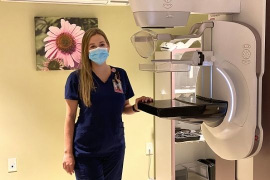
3D Mammography
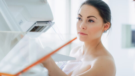
All mammography exams are performed using our 3D digital mammography systems.
3D mammography, or breast tomosynthesis, is the latest advance in breast imaging. Like traditional mammography, 3D mammography uses X-rays to produce images of breast tissue in order to detect lumps, tumors or other abnormalities. 3D mammography is capable of producing more detailed images of breast tissue.
3D captures multiple slices of the breast, all at different angles. The images are brought together to create crystal clear 3D reconstruction of the breast. The radiologist is then able to review reconstruction, one slide at a time, almost like turning pages in a book. This makes it easier for doctors to see if there’s anything to be concerned about.

3D Automated Breast Ultrasound (ABUS)
More than 40% of all women have “dense breasts”. Dense breast tissue can hide breast cancer and is a significant risk factor for getting breast cancer. ABUS is an innovative breast cancer screening for women with dense breast tissue. Mammography may miss over 1/3 of cancers in dense breasts. The InveniaTM ABUS (Automated Breast Ultrasound System) is a comfortable, radiation free, alternative to other supplemental screening options for women with dense breast tissue. When used in addition to mammography, Invenia ABUS can improve breast cancer detection by 55 percent over mammography alone.
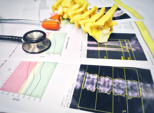
Bone Density Testing (DEXA)
Bone density testing (DEXA) determines if you have osteoporosis — which can cause bones to become more fragile and more likely to break
In the past, osteoporosis could be detected only after you broke a bone. By that time, however, your bones could be quite weak. A bone density test makes it possible to know your risk of breaking bones before the fact. A bone density test uses X-rays to measure how many grams of calcium and other bone minerals are packed into a segment of bone. The bones that are most commonly tested are located in the spine, hip and forearm.
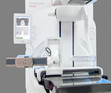
Breast Biopsy Procedures
3D Stereo Guided Breast Biopsy : In keeping with our history of leading the way in our field, CBIS is proud to have the most advanced biopsy system available. We now use the GE Healthcare Senographe Pristina “Serena” 3D stereotactic biopsy system. This system allows us to use tomosynthesis (3D mammography) to precisely locate abnormalities, especially those that were best (or only) seen on 3D mammography. Biopsies done this way use less radiation and are completed more quickly and accurately than with previous methods.
A breast biopsy involves removing a sample of breast tissue to determine whether it is cancerous or benign (non-cancerous). While physical breast exam, mammography, ultrasound, and other breast imaging methods can help detect a breast abnormality, biopsy followed by pathological (microscopic) analysis of the breast biopsy result is the only definitive way to determine if cancer is present.
Once a sample of breast tissue has been removed by the radiologist, it is sent to the laboratory for microscopic examination by a pathologist. A clinical pathologist is a physician who performs laboratory analyses of tissues to determine their type.
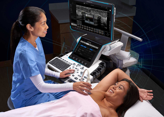
Diagnostic Breast Ultrasound
Breast ultrasound, also known as sonography or ultrasonography, is frequently used to evaluate breast abnormalities that are found with screening or diagnostic mammography, screening ultrasound or during self exam or a physician performed clinical breast exam
Ultrasound allows significant freedom in obtaining images of the breast from almost any orientation. Ultrasound is excellent at imaging cysts: round, fluid-filled, pockets inside the breast. Additionally, ultrasound can often quickly determine if a suspicious area is, in fact, a cyst (always non-cancerous) or an increased density of solid tissue (dense mass) which may require a biopsy to determine if it is malignant (cancerous).
Contrast Enhanced Mammography
Contrast Enhanced Spectral Mammography procedure is one of more recent advances in early breast cancer detection. Dr. Schroeder is one of the leading pioneers of this technique and has been using this tool since its approval in 2012 when he was able to bring CESM to Greenville making it the second city in the U.S. after Beverly Hills, CA. This innovative tool is rapidly growing in popularity as a low cost, comfortable, accurate alternative to breast MRI.
Contrast Enhanced Spectral Mammography (CESM) is a special type of mammogram that is performed after an IV injection of X-ray contrast (iodine). CESM shows all of the information of a regular mammogram but also shows areas of increased blood supply. Breast cancer typically has a greater blood supply than normal tissue so it is highlighted on the images.
Ductography
Ductography (also called galactography) is a special type of contrast-enhanced mammography used for imaging the breast ducts. Ductography can aid in diagnosing the cause of an abnormal nipple discharge and is valuable in diagnosing breast cancer, intraductal papillomas and other conditions. Papillomas are wart-like, non-cancerous tumors with branchings or stalks that have grown inside the breast duct; they are the most common cause of nipple discharge.
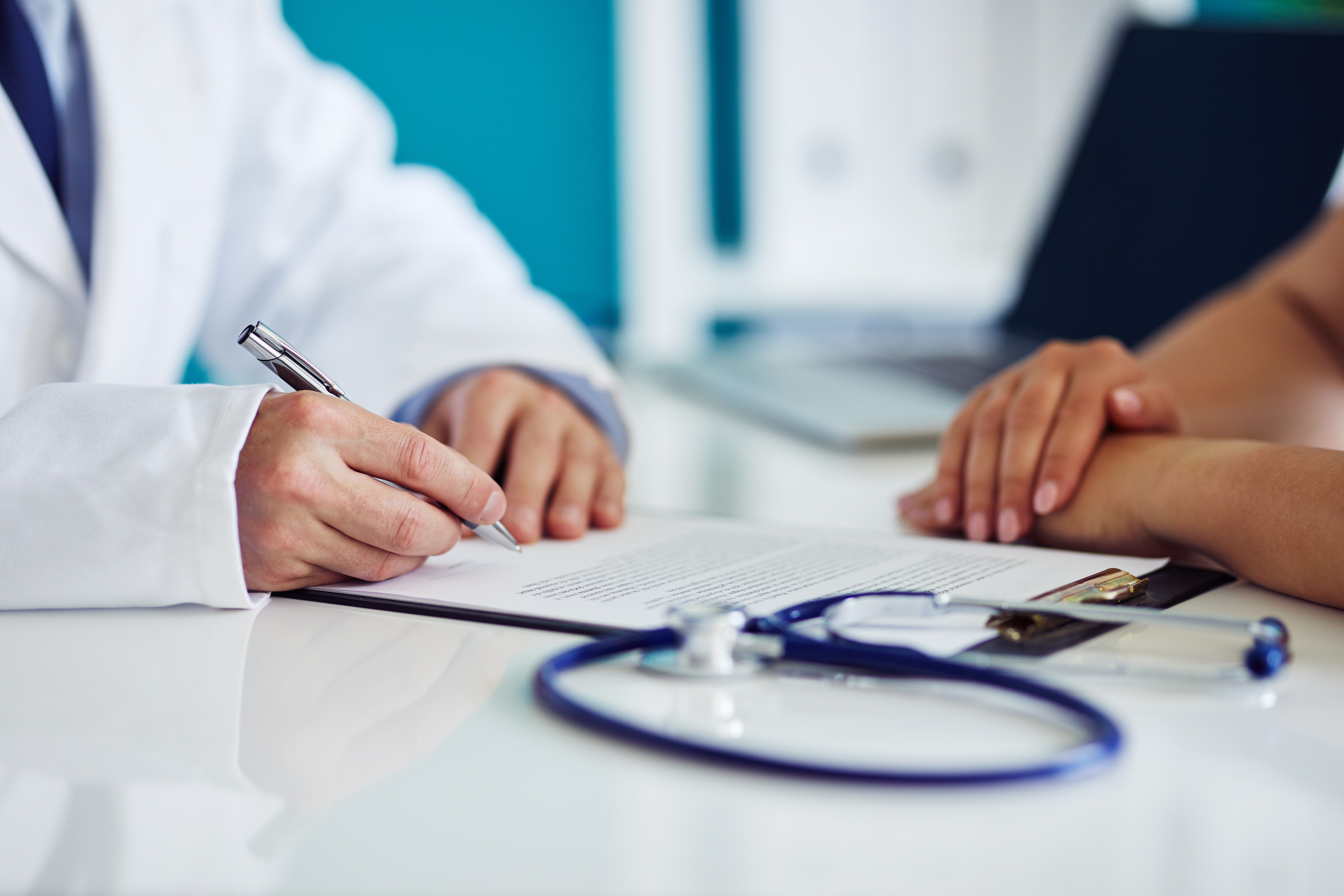
Second Opinion Consultations
In addition to our wide range of breast imaging services, our specialized expert breast imaging radiologists also offer second opinion consultations. As with other second opinion consultation services, your initial tests were completed at another facility, but you may prefer a specialist’s opinion of the images or review of the findings.
Our radiologist provides a comprehensive review and analysis of your patient information and test results. After an initial consultation, we will determine if additional imaging or procedures are required to establish a clear opinion and diagnosis. We also work with patients to develop a strategic plan based on your personal information, test results, and family history. Research has shown that nearly half of all patients who were told they needed a breast biopsy by non-specialist radiologists did not need a biopsy at all. If you have received an abnormal breast imaging exam and are told you need a biopsy we will provide our second opinion at no charge.